LUYOR-3109高強度紫外催化光源促銷
LUYOR-3109紫外光源采用了9顆365nm大功率led,安裝有二次光學透鏡,輸出紫外線強度高,...
2024-08-08作者:激發光源事業部時間:2019-09-05 10:26:27瀏覽18761 次
mCherry是一種來自于蘑菇珊瑚(mushroom coral)的紅色熒光蛋白,常有于標記和示蹤某些分子和細胞組分。相對于其他熒光,mCherry的好處在于它的顏色和應用最多的綠色熒光蛋白(GFP)能進行共同標記,并且mCherry相對于其他單體熒光蛋白來說也具有卓越的光穩定型。
美國路陽生產一款便攜式熒光蛋白激發光源,能夠方便快捷觀察EGFP在動植物體內的表達。我們免費提供樣機供科研院所試用,滿意付款。如有興趣,可添加微信號(luyor01)聯系。
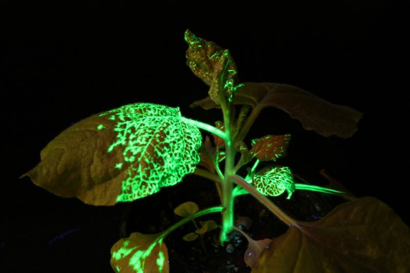
EGFP在煙草上的表達
綠色熒光蛋白在果蠅上的表達
mcherry Fluorescent Proteins Excitation Emission
mCherry是一種來自于蘑菇珊瑚(mushroom coral)的紅色熒光蛋白,常有于標記和示蹤某些分子和細胞組分。相對于其他熒光,mcherry的好處在于它的顏色和應用最多的綠色熒光蛋白(GFP)能進行共同標記,并且mcherry相對于其他單體熒光蛋白來說也具有卓越的光穩定型。
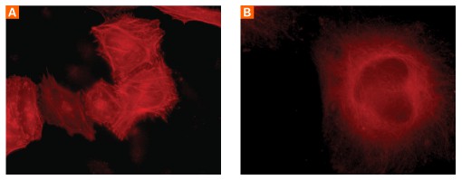
吸收和發射波長:
mcherry的更大吸收/發射峰分別位于587nm和610nm,對光致漂白耐受,熒光非常穩定。

?mCherry常用于與目的基因組成融合蛋白以及通過IRES或2A與感興趣的蛋白共表達;
?啟動子活性研究;
?熒光共振能量轉移(fluorescence resonance energy transfer,FRET)和其他定量實驗;
?標記細胞或者分子,進行示蹤實驗;
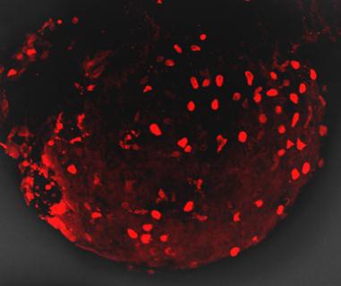
mCherry is a fluorophore (a fluorescent protein) used in biotechnology as a tracer to follow the flow of fluids, as a marker when tagged to molecules and cell components. mCherry and the majority of red fluorescent proteins derive from a protein isolated from Discosoma sp., while other fluorescent proteins in the green range are often variants of GFP from Aequorea victoria.
m前綴的意義:
mCherry的m為monmer單體的縮寫,表示mCherry熒光蛋白的形式為單體,這在很多實驗設計中非常重要,比如與目的標記基因組成融合蛋白時。
成熟時間:
mCherry具有較快的成熟速度,t0.5為15分鐘,這在一些需要做出快速反應的實驗非常重要。比如啟動子活性報告系統等。
你可以選用LUYOR-3415RG和LUYOR-3260GR便攜式熒光蛋白激發光源來直接觀察和篩選mcherry在植物中有沒有表達。如需進一步了解,請撥打電話153-1756-5658進行咨詢(或直接添加微信15317565658咨詢)。
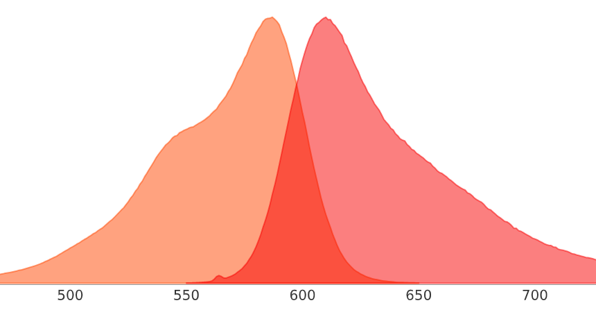
mcherry紅色熒光蛋白的激發波長和發射波長
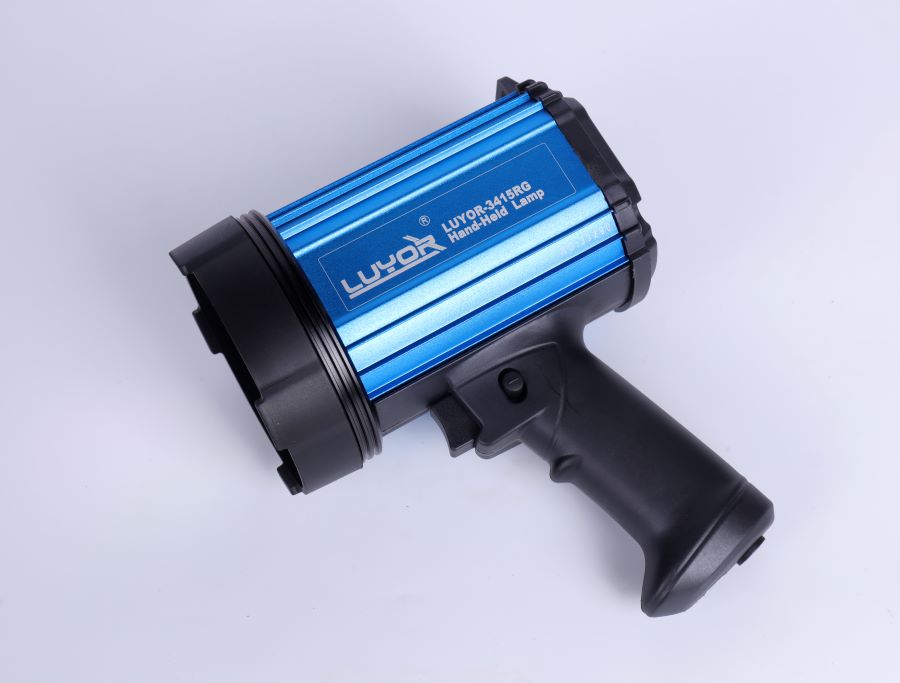
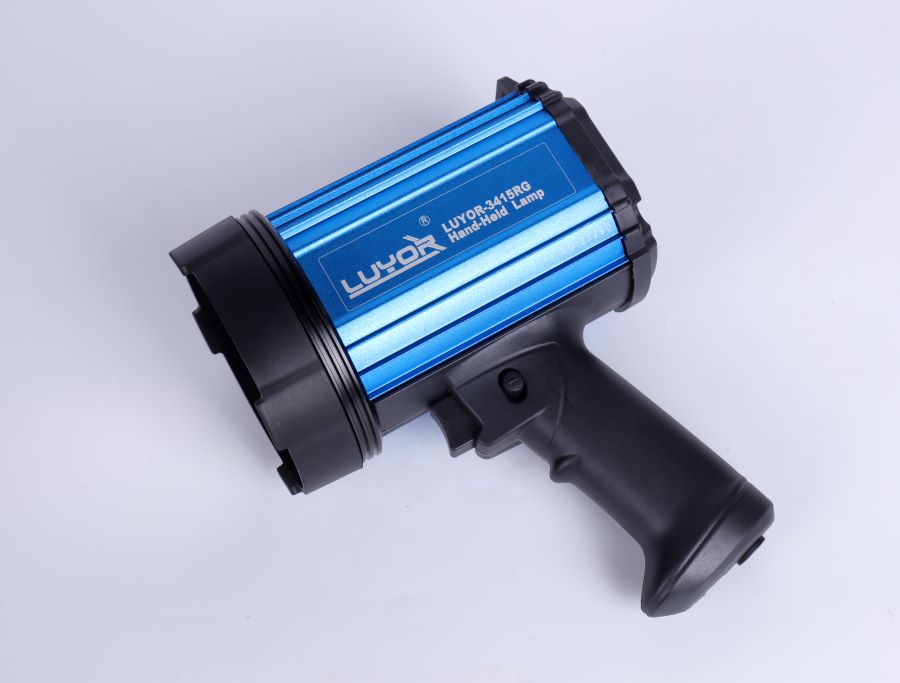
上圖為LUYOR-3415RG便攜式紅色熒光蛋白激發光源
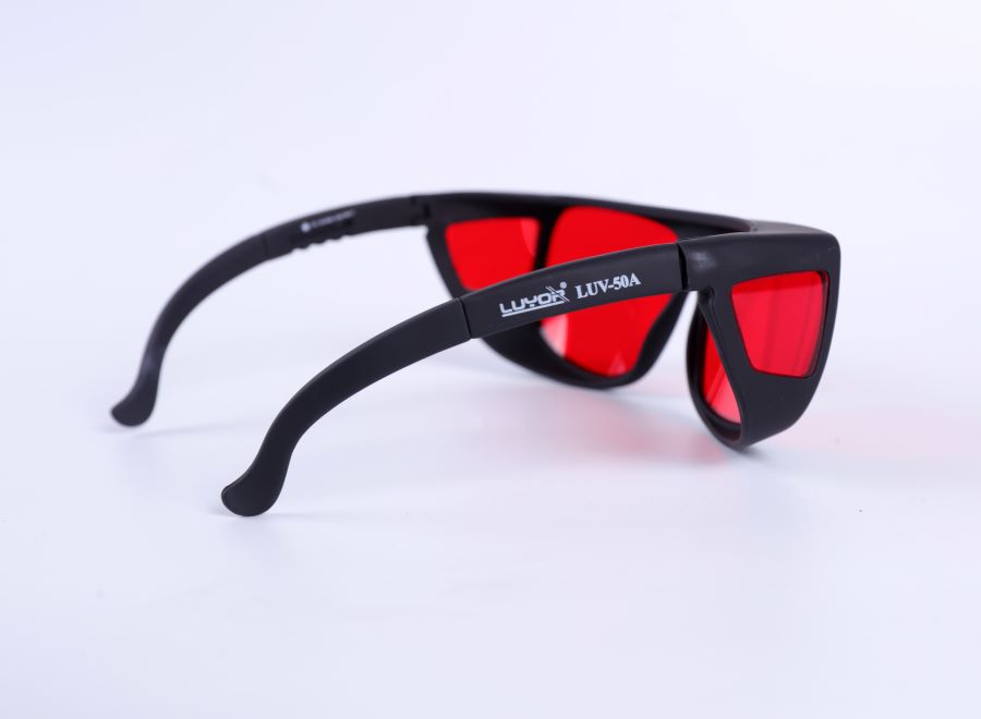
上圖為LUV-50A紅色熒光蛋白觀察眼鏡
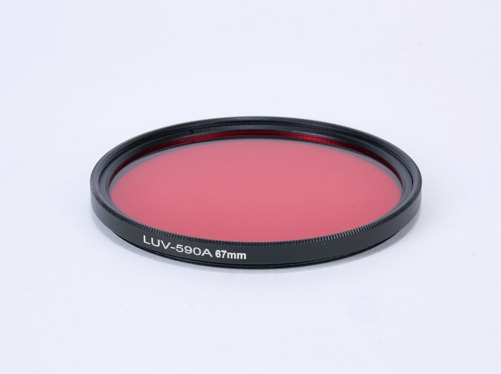
上圖為LUV-590A 紅色熒光蛋白拍照濾鏡
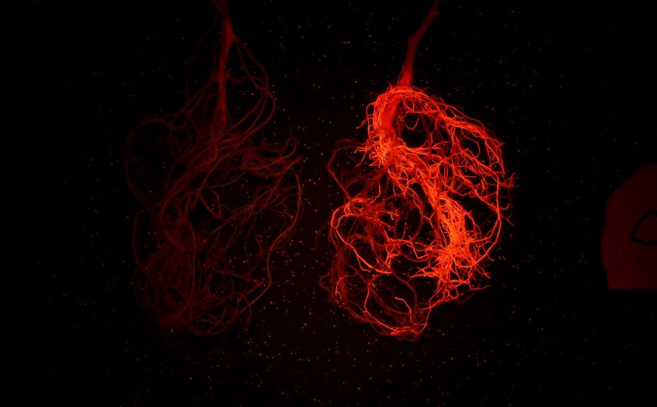
上圖為紅色熒光蛋白在大豆根系上的表達(LUYOR-3415RG照射,LUV-590A濾鏡拍攝)
紫外熒光蛋白UV Proteins
| Protein | Excitation Wavelength | Emission Wavelength |
|---|---|---|
| Sirius | 355 | 424 |
| Sandercyanin | 375 | 630 |
| shBFP-N158S/L173I | 375 | 458 |
藍色熒光蛋白Blue Proteins
| Protein | Excitation Wavelength | Emission Wavelength |
|---|---|---|
| Azurite | 383 | 450 |
| EBFP2 | 383 | 448 |
| mKalama1 | 385 | 456 |
| mTagBFP2 | 399 | 454 |
| TagBFP | 402 | 457 |
| shBFP | 401 | 458 |
青色熒光蛋白Cyan Proteins
| Protein | Excitation Wavelength | Emission Wavelength |
|---|---|---|
| ECFP | 433 | 475 |
| Cerulean | 433 | 475 |
| mCerulean3 | 433 | 475 |
| SCFP3A | 433 | 474 |
| CyPet | 435 | 477 |
| mTurquoise | 434 | 474 |
| mTurquoise2 | 434 | 474 |
| TagCFP | 458 | 480 |
| mTFP1 | 462 | 492 |
| monomeric Midoriishi-Cyan | 470 | 496 |
| Aquamarine | 430 | 474 |
綠色熒光蛋白Green Proteins
| Protein | Excitation Wavelength | Emission Wavelength |
|---|---|---|
| TurboGFP | 482 | 502 |
| TagGFP2 | 483 | 506 |
| mUKG | 483 | 499 |
| Superfolder GFP | 485 | 510 |
| Emerald | 487 | 509 |
| EGFP | 488 | 507 |
| Monomeric Azami Green | 492 | 505 |
| mWasabi | 493 | 509 |
| Clover | 505 | 515 |
| mNeonGreen | 506 | 517 |
| NowGFP | 494 | 502 |
| mClover3 | 506 | 518 |
黃色熒光蛋白Yellow Proteins
| Protein | Excitation Wavelength | Emission Wavelength |
|---|---|---|
| TagYFP | 508 | 524 |
| EYFP | 513 | 527 |
| Topaz | 514 | 527 |
| Venus | 515 | 528 |
| SYFP2 | 515 | 527 |
| Citrine | 516 | 529 |
| Ypet | 517 | 530 |
| lanRFP-ΔS83 | 521 | 592 |
| mPapaya1 | 530 | 541 |
| mCyRFP1 | 528 | 594 |
桔色熒光蛋白Orange Proteins
| Protein | Excitation Wavelength | Emission Wavelength |
|---|---|---|
| Monomeric Kusabira-Orange | 548 | 559 |
| mOrange | 548 | 562 |
| mOrange2 | 549 | 565 |
| mKOκ | 551 | 563 |
| mKO2 | 551 | 565 |
紅色熒光蛋白Red Proteins
| Protein | Excitation Wavelength | Emission Wavelength |
|---|---|---|
| TagRFP | 555 | 584 |
| TagRFP-T | 555 | 584 |
| RRvT | 556 | 583 |
| mRuby | 558 | 605 |
| mRuby2 | 559 | 600 |
| mTangerine | 568 | 585 |
| mApple | 568 | 592 |
| mStrawberry | 574 | 596 |
| FusionRed | 580 | 608 |
| mCherry | 587 | 610 |
| mNectarine | 558 | 578 |
| mRuby3 | 558 | 592 |
| mScarlet | 569 | 594 |
| mScarlet-I | 569 | 593 |
遠紅熒光蛋白Far Red Proteins
| Protein | Excitation Wavelength | Emission Wavelength |
|---|---|---|
| mKate2 | 588 | 633 |
| HcRed-Tandem | 590 | 637 |
| mPlum | 590 | 649 |
| mRaspberry | 598 | 625 |
| mNeptune | 600 | 650 |
| NirFP | 605 | 670 |
| TagRFP657 | 611 | 657 |
| TagRFP675 | 598 | 675 |
| mCardinal | 604 | 659 |
| mStable | 597 | 633 |
| mMaroon1 | 609 | 657 |
| mGarnet2 | 598 | 671 |
近紅熒光蛋白Near IR Proteins
| Protein | Excitation Wavelength | Emission Wavelength |
|---|---|---|
| iFP1.4 | 684 | 708 |
| iRFP713 (iRFP) | 690 | 713 |
| iRFP670 | 643 | 670 |
| iRFP682 | 663 | 682 |
| iRFP702 | 673 | 702 |
| iRFP720 | 702 | 720 |
| iFP2.0 | 690 | 711 |
| mIFP | 683 | 704 |
| TDsmURFP | 642 | 670 |
| miRFP670 | 642 | 670 |
Sapphire-type Proteins
| Protein | Excitation Wavelength | Emission Wavelength |
|---|---|---|
| Sapphire | 399 | 511 |
| T-Sapphire | 399 | 511 |
| mAmetrine | 406 | 526 |
Red fluorescent proteins (RFP) can be imaged on existing confocal or widefield microscopes, and they also have more penetrating power. The excitation and emission maxima of RFP are 558nm and 583 nm, respectively.
The use of RFP, however, has been hampered with several issues. RFP is an obligate tetramer - thus, it forms large aggregates inside cells. This makes the use to RFP to report the location of a protein severely limited.
Although GFP can successfully fuse with several hundreds of proteins, RFP-conjugated proteins are often toxic. Some variants of RFP have overcome these limitations. For example, DsRed2 fluorescent protein does not form aggregates and has reduced toxicity, while another variant of RFP (known as RedStar) has increased brightness and maturation rate.